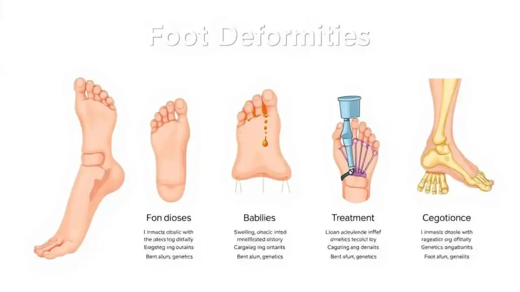
Understanding Foot Deformities:Causes,Symptoms,and Treatments
Our feet are remarkable structures, intricate masterpieces of engineering that bear the brunt of our daily activities, providing balance, propulsion, and shock absorption. We often take their complex functionality for granted until something goes awry. Foot deformities, whether congenital or acquired, can significantly impact our mobility, cause chronic pain, and diminish our overall quality of life. Understanding these conditions is the first step toward effective management and regaining comfortable movement.
As we delve into the world of foot deformities, we aim to unravel some of the most common conditions we encounter: Equinus, Calcaneus, Varus, Valgus, Cavus, Planus, and Splay. For each, we will explore its definition, the underlying causes, the tell-tale symptoms, and the range of available treatment options.
“The foot is the foundation upon which the entire body stands. When its intricate balance is disrupted, the effects can ripple throughout the kinetic chain, impacting everything from gait to spinal alignment.” — A.J. McInnes, Orthopaedic Surgeon
Let’s explore these conditions in detail.
1. Equinus Deformity
What it is: Equinus describes a condition where the ankle lacks the ability to adequately dorsiflex, meaning the foot cannot be pulled upwards towards the shin beyond a neutral 90-degree angle. This results in the foot being perpetually pointed downwards, resembling a horse’s hoof (hence “equinus”).
Why it happens:
- Congenital factors: Short Achilles tendon (tight heel cord) present from birth.
- Neurological conditions: Cerebral palsy, stroke, spina bifida, or polio, which can lead to muscle spasticity or paralysis.
- Idiopathic: No clear identifiable cause.
- Trauma: Injuries that lead to scar tissue formation or improper healing.
- Improper footwear: Prolonged use of high-heeled shoes can shorten the Achilles tendon over time.
What you might feel:
- Difficulty walking, often leading to walking on the toes or forefoot (toe-walking).
- Compensatory bending of the knee or hip to allow the foot to clear the ground.
- Foreshortened stride and increased pressure on the ball of the foot.
- Formation of calluses or corns under the ball of the foot or on the toes.
- Associated conditions like hammertoes, bunions, or metatarsalgia due to altered weight distribution.
What can be done:
- Conservative:
- Stretching exercises, especially for the calf muscles and Achilles tendon.
- Night splints or braces to maintain ankle dorsiflexion.
- Physical therapy to improve range of motion and gait.
- Orthotics to support the foot and redistribute pressure.
- Surgical:
- Achilles tendon lengthening (gastroc recession or Achilles fractional lengthening) to improve ankle dorsiflexion.
2. Calcaneus Deformity
What it is: The opposite of equinus, a calcaneus deformity involves excessive dorsiflexion of the ankle, meaning the foot is permanently pointed upwards, with the heel touching or nearly touching the ground. This often results in weight being borne primarily on the heel.
Why it happens:
- Muscle Weakness/Paralysis: Damage to the calf muscles or the nerves controlling them (e.g., from spina bifida, polio, or nerve injury).
- Congenital: Less common than equinus, but can be present at birth.
- Trauma: Injuries to the ankle joint or surrounding tendons that lead to imbalance.
What you might feel:
- Difficulty pushing off the ground during walking due to absent or weak plantarflexion.
- Increased pressure and potential pain on the heel.
- A flat-footed appearance, but distinct from pes planus as it’s primarily an ankle position issue.
- Compensatory postural changes, often affecting the knees and hips.
What can be done:
- Conservative:
- Physical therapy to strengthen weakened muscles and improve balance.
- Ankle-foot orthoses (AFOs) to provide support and limit excessive dorsiflexion.
- Custom footwear to accommodate the foot’s position and provide cushioning for the heel.
- Surgical:
- Tendon transfers or fusions to stabilize the ankle and restore muscle balance.
3. Varus Deformity
What it is: A varus deformity refers to an inward angulation of a body part. In the foot, this typically means the heel (hindfoot varus) or the forefoot (forefoot varus) turns inwards, causing the person to walk on the outer edge of their foot. This can lead to an increased arch height or a supinated foot posture.
Why it happens:
- Congenital: Clubfoot (Talipes Equinovarus) is a common congenital condition involving multiple deformities, including hindfoot varus.
- Neurological conditions: Muscle imbalances due to conditions like Charcot-Marie-Tooth disease or spasticity.
- Idiopathic: Can develop without a clear cause.
- Trauma or arthritis: Damage to the joints or ligaments can lead to instability and inward turning.
What you might feel:
- Pain along the outer edge of the foot and ankle.
- Increased incidence of ankle sprains due to instability.
- Formation of calluses or corns on the lateral (outer) side of the foot.
- Difficulty finding comfortable shoes.
- A “bowed leg” appearance if the varus extends up the leg.
What can be done:
- Conservative:
- Orthotics with lateral wedges or posts to support the foot and redistribute pressure.
- Physical therapy to strengthen ankle stabilizers and improve proprioception.
- Supportive footwear with good ankle support.
- Surgical:
- Osteotomies (bone cuts) to realign the foot and ankle bones.
- Tendon transfers to balance muscle forces.
- Arthrodesis (joint fusion) for severe, rigid deformities.
4. Valgus Deformity
What it is: A valgus deformity is the opposite of varus, involving an outward angulation of a body part. In the foot, this means the heel (hindfoot valgus) or forefoot (forefoot valgus) turns outwards, causing the person to walk on the inner edge of their foot. This is often associated with a flattened arch (pes planus).
Why it happens:
- Ligamentous laxity: “Loose” ligaments that fail to adequately support the arches, especially in children.
- Muscle imbalance: Weakness in supporting muscles or tightness in opposing muscles.
- Obesity: Increased weight puts more strain on the arches.
- Arthritis or trauma: Damage to joints or ligaments, particularly the posterior tibial tendon.
- Congenital: Can be part of a broader syndrome or isolated.
What you might feel:
- Pain along the inner arch and ankle.
- Increased pressure areas on the medial (inner) side of the foot.
- Overpronation during gait, leading to internal rotation of the leg.
- Associated conditions like bunions (hallux valgus) due to increased pressure on the big toe joint.
- A “knock-knee” appearance if the valgus extends up the leg.
What can be done:
- Conservative:
- Custom orthotics with medial arch support to control pronation.
- Supportive shoes with a firm heel counter.
- Physical therapy to strengthen intrinsic foot muscles and improve gait mechanics.
- Weight management for obese individuals.
- Surgical:
- Tendon repairs or transfers (e.g., posterior tibial tendon repair).
- Osteotomies to realign bones.
- Fusions for severe, rigid flatfoot deformities.
5. Cavus Foot (Pes Cavus)
What it is: A cavus foot, or pes cavus, is characterized by an abnormally high arch that does not flatten when weight is borne. This results in the foot primarily bearing weight on the heel and the ball of the foot, with minimal contact from the midfoot.
Why it happens:
- Neurological conditions: The most common cause, including Charcot-Marie-Tooth disease, spina bifida, cerebral palsy, or poliomyelitis.
- Inherited: Can run in families without a clear neurological cause.
- Idiopathic: Some cases have no identifiable cause.
What you might feel:
- Pain in the heel and ball of the foot due to concentrated pressure.
- Hammertoes or claw toes due to muscle imbalance.
- Calluses or corns under the metatarsal heads (ball of the foot) and on the toes.
- Frequent ankle sprains due to the foot’s supinated (outwardly rotated) position.
- Difficulty finding shoes that comfortably accommodate the high instep.
- Reduced shock absorption, leading to knee, hip, and back pain.
What can be done:
- Conservative:
- Custom orthotics to redistribute pressure, provide cushioning, and stabilize the foot.
- Supportive, well-cushioned shoes with a deep toe box.
- Stretching exercises for tight muscles (e.g., Achilles tendon, plantar fascia).
- Padding for calluses and corns.
- Surgical:
- Osteotomies to rebalance the foot bones.
- Tendon transfers to improve muscle balance.
- Soft tissue releases for tight structures.
- Arthrodesis (fusion) for severe, rigid deformities, especially in cases of progressive neurological disease.
6. Planus Foot (Pes Planus)
What it is: A planus foot, commonly known as a flat foot, is characterized by a collapsed or very low arch. In many cases, the entire sole of the foot makes contact with the ground when standing. Flat feet can be flexible (arch reappears when non-weight-bearing) or rigid (arch remains flat even when non-weight-bearing).
Why it happens:
- Genetics: Often inherited from family members.
- Ligamentous laxity: “Loose” ligaments that stretch easily, failing to maintain the arch shape.
- Posterior Tibial Tendon Dysfunction (PTTD): Weakening or tearing of the posterior tibial tendon, which is crucial for supporting the arch.
- Rheumatoid arthritis: Inflammatory arthritis can damage foot joints and ligaments.
- Obesity: Increased weight strains the arches.
- Injury: Trauma to ankle or foot bones/ligaments.
- Aging: Arches can flatten over time.
What you might feel:
- Pain along the inner side of the ankle and arch.
- Fatigue in the feet and legs, especially after prolonged standing or walking.
- Swelling along the inside of the ankle.
- Pain in the knee, hip, or lower back due to altered gait mechanics.
- Increased risk of bunions, hammertoes, and Achilles tendonitis.
- Difficulty with specific sports or activities requiring stability.
What can be done:
- Conservative:
- Custom orthotics to support the arch and control pronation.
- Supportive shoes with a firm heel counter and motion control features.
- Physical therapy to strengthen arch-supporting muscles and improve flexibility.
- Weight management.
- Rest, ice, compression, and elevation (RICE) for acute pain.
- Surgical: (Primarily for severe, painful, or rigid flatfoot not responding to conservative care)
- Tendon grafts or transfers.
- Osteotomies to reshape bones.
- Arthrodesis (fusion) for severe, fixed deformities.
7. Splay Foot
What it is: Splay foot refers to a condition where the forefoot (the front part of the foot) widens and spreads out, often leading to increased space between the toes, particularly the first and second toes. This widening can result in the metatarsal bones spreading abnormally. It is often associated with the development of bunions (hallux valgus).
Why it happens:
- Ligamentous laxity: Weakness in the transverse ligaments that hold the metatarsal heads together.
- Genetics: Predisposition to looser connective tissues.
- Improper footwear: Narrow or pointed-toe shoes can exacerbate forefoot spreading over time.
- Excessive pronation: Overpronation can put outward pressure on the forefoot.
- Muscle imbalance: Weakness in the intrinsic foot muscles.
- Aging: Degenerative changes can lead to overall foot widening.
What you might feel:
- Pain across the ball of the foot (metatarsalgia), especially under the second and third toes.
- Developing bunions (hallux valgus) due to the big toe drifting outwards.
- Splaying of the toes, making them appear “spread out.”
- Difficulty finding shoes that are wide enough, even in regular sizes.
- Calluses or corns under the metatarsal heads.
- Hammer toes or claw toes.
What can be done:
- Conservative:
- Wide-fitting shoes with a spacious toe box.
- Orthotics with metatarsal pads or domes to support the transverse arch.
- Toe spacers to help realign toes and reduce friction.
- Physical therapy to strengthen intrinsic foot muscles.
- Avoiding high heels or narrow shoes.
- Surgical: (Typically reserved for symptomatic bunions or severe splay causing significant pain/deformity)
- Bunionectomy (hallux valgus correction).
- Metatarsal osteotomies to narrow the forefoot.
Summary of Foot Deformities
To provide a quick reference, here’s a summary of the foot deformities we’ve discussed:


