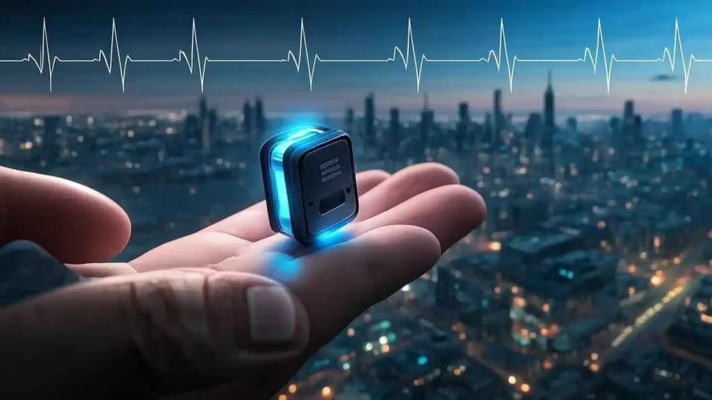
The Dawn of a New Era: Understanding the Revolutionary Rice-Sized Pacemaker
A cardiac pacemaker is a small, battery-powered device that is implanted in the chest to help regulate the heartbeat. It consists of a pulse generator and one or more electrodes, which are small wires that connect the generator to the heart. The generator produces electrical impulses, which are delivered through the electrodes to stimulate the heart muscle and maintain a regular heart rhythm.
There are several types of pacemakers, but the most common one is the single-chamber pacemaker, which has one electrode placed in the right atrium or right ventricle of the heart. Dual-chamber pacemakers have two electrodes, one in the right atrium and one in the right ventricle, allowing for better coordination between the two chambers. Biventricular pacemakers are used for patients with heart failure, and they have three electrodes, one in each ventricle and one in the right atrium.
Pacemakers are used to treat various heart rhythm disorders, including bradycardia (slow heart rate), tachycardia (fast heart rate), and atrial fibrillation (irregular heart rhythm). Bradycardia can be caused by damage to the heart’s electrical system, certain medications, or age-related changes in the heart’s conduction system. Tachycardia can result from heart disease, high blood pressure, or other medical conditions. Atrial fibrillation is a common heart rhythm disorder that affects millions of people worldwide and can lead to stroke, heart failure, and other complications.
The decision to implant a pacemaker is based on a thorough evaluation of the patient’s medical history, symptoms, physical examination, and diagnostic tests, such as an electrocardiogram (ECG), echocardiogram, and stress test. The procedure to implant a pacemaker is usually done under local anesthesia and takes about one to two hours. The patient typically stays in the hospital for one to two days after the procedure to recover and undergo monitoring.
Once implanted, the pacemaker continuously monitors the heart rate and adjusts the electrical impulses accordingly to maintain a regular heart rhythm. The battery life of a pacemaker can last up to 10 years, after which it needs to be replaced through a minor surgical procedure.
A cardiac pacemaker is a life-saving device that helps regulate the heartbeat in patients with various heart rhythm disorders. It works by generating electrical impulses that stimulate the heart muscle and maintain a regular heart rhythm. The decision to implant a pacemaker is based on a thorough evaluation of the patient’s medical history and symptoms, and the procedure is usually done under local anesthesia with a short recovery period.
What Exactly is a Pacemaker? A Closer Look at This Life-Saving Device
At its very core, a pacemaker is a sophisticated, implanted medical device meticulously designed to regulate the heart’s rhythm. It serves as a vital intervention, primarily used to treat various types of arrhythmias, particularly those characterized by an abnormally slow heart rate, known medically as bradycardia, or significant blocks in the heart’s natural electrical conduction system.
Imagine your heart as a finely tuned orchestra, and its natural electrical impulses are the conductor, ensuring each beat occurs at the right moment and with the correct rhythm. When this natural conduction system falters, leading to slow or irregular beats, the heart may not pump enough oxygenated blood to the body, causing symptoms like fatigue, dizziness, shortness of breath, or even fainting. This is where the pacemaker steps in, acting as a tiny, internal electrician. It stands ready to monitor the heart’s activity and, when necessary, send out precise, low-energy electrical signals to stimulate the heart muscle, ensuring beats occur consistently and at an appropriate, healthy pace.
A typical pacemaker system, though small and discreet once implanted, consists of two essential and interconnected main parts:
- The Pulse Generator: This is the brains and power source of the system. It’s a small, hermetically sealed metal box, often no larger than a pocket watch or a large silver dollar, and typically weighing just an ounce or two. It’s usually implanted just beneath the skin, most commonly in the upper chest near the collarbone. Inside this durable casing are two critical components:
- The Battery: A long-lasting battery, specially designed for medical implants, powers the device. Modern pacemaker batteries are incredibly efficient and can last anywhere from 5 to 15 years, depending on the individual’s pacing needs.
- The Electronic Circuitry: This incredibly sophisticated micro-circuitry is responsible for continuously monitoring the heart’s intrinsic electrical rhythm. It processes this information using complex algorithms, identifies abnormalities, and then determines precisely when and how to generate the minute electrical pulses needed to correct the rhythm.
- One or More Leads (or Wires): These are thin, highly flexible, insulated wires that are carefully guided through a vein (usually in the shoulder area) directly into the heart muscle tissue within its chambers. Depending on the patient’s specific condition, a pacemaker system might have one, two, or even three leads, targeting different chambers of the heart (e.g., the right atrium, the right ventricle, or both, and sometimes the left ventricle for specialized therapies). These leads perform two absolutely vital, bidirectional functions:
- Sensing: They cleverly detect the heart’s own natural electrical activity, transmitting this critical information back to the pulse generator. This allows the pacemaker to “listen” to the heart and only deliver a pulse when it’s truly needed, optimizing battery life and preventing unnecessary interventions.
- Pacing: When the pulse generator determines that a beat is missed or too slow, the leads transmit the precisely timed electrical pulses from the generator directly to the heart muscle, stimulating it to contract and initiate a heartbeat. The very tip of the lead often has a tiny screw or barb that gently anchors it into the heart wall to ensure stable, reliable contact.
Over the decades, pacemakers have undergone a profound and often revolutionary evolution. Early models, first clinically used in the late 1950s and early 1960s, were comparatively bulky, sometimes external devices that limited patient mobility. In stark contrast, modern pacemakers are astonishingly miniaturized, exceptionally durable, and incredibly advanced, boasting an array of sophisticated features that vastly improve patient quality of life and clinical outcomes. These advancements include:
- Rate Responsiveness: This critical feature allows the pacemaker to intelligently adjust the heart rate based on the patient’s physical activity level. For example, it can gently increase the heart rate during exercise and slow it down during rest or sleep, mimic the body’s natural response to exertion.
- Remote Monitoring: Many contemporary pacemakers can wirelessly transmit data to a patient’s clinic, allowing healthcare providers to monitor the device’s function and the heart’s rhythm from a distance, reducing the need for frequent in-person appointments and enabling early detection of potential issues.
- MRI Compatibility: Newer models are designed to be safe for Magnetic Resonance Imaging (MRI) scans, which was once a major contraindication for pacemaker patients, significantly expanding diagnostic options.
- Leadless Pacemakers: A groundbreaking innovation, these tiny devices (about the size of a large vitamin) are implanted directly into the heart without the need for traditional leads, further reducing potential complications.
- Diagnostic Capabilities: Modern pacemakers also collect and store valuable data about arrhythmias and heart function, providing physicians with detailed insights to guide ongoing treatment.
In essence, the pacemaker has transformed from a life-sustaining device into a sophisticated, highly adaptable heart rhythm management system, offering patients with specific cardiac conditions the ability to lead full, active, and significantly improved lives.
Here’s a simple breakdown of the components:
| Component | Function |
| Pulse Generator | Contains battery and circuitry; monitors heart, generates electrical pulses. |
| Leads (Wires) | Transmit pulses to heart; sense heart’s natural electrical activity. |
| Battery | Powers the pulse generator; typically lasts 7-15 years. |
| Electronic Logic | Interprets signals, decides when and how to pace. |
How Does a Pacemaker Work? The Intricate Dance of Electricity
The human heart is an astonishingly complex organ, a muscular pump that beats tirelessly, distributing oxygen and nutrients throughout the body. Its rhythmic contraction is orchestrated by a precise internal electrical system, a symphony of impulses that ensures every beat is perfectly timed. When this natural rhythm falters, an artificial pacemaker steps in, acting as an intelligent conductor to restore the heart’s harmonious “dance of electricity.”
The Heart’s Natural Conductor: The Intrinsic Electrical System
To truly appreciate the wonder of a pacemaker, we must first understand the heart’s innate electrical architecture. Far from being a mere muscle, the heart contains specialized cells capable of generating and conducting electrical signals:
- The Sinoatrial (SA) Node – The Heart’s Primary Pacemaker: Located in the top right chamber of the heart (the right atrium), the SA node is often referred to as the heart’s natural pacemaker. It’s a small cluster of specialized cells with the remarkable ability to spontaneously generate electrical impulses at a regular, intrinsic rate, typically between 60 to 100 beats per minute at rest. These impulses are the very spark that initiates each heartbeat.
- Atrial Contraction: Once generated by the SA node, these electrical impulses swiftly spread like ripples across both the right and left atria. This rapid propagation causes the atrial muscle cells to contract, pushing blood from the atria down into the ventricles (the lower chambers) in preparation for the next phase of pumping.
- The Atrioventricular (AV) Node – The Crucial Gateway: The electrical signal then arrives at the atrioventricular (AV) node, a cluster of cells situated at the junction between the atria and the ventricles. The AV node acts as a vital “gateway” or a “damper.” It intentionally delays the electrical signal for a critical fraction of a second. This brief delay is crucial, allowing the ventricles sufficient time to fully fill with blood from the contracting atria before they, in turn, contract. Without this delay, the pumping action would be inefficient.
- Ventricular Conduction System – The Rapid Distribution Network: After passing through the AV node, the signal travels down through specialized conductive pathways:
- Bundle of His: This pathway emerges from the AV node and quickly branches into two main divisions.
- Bundle Branches (Left and Right): These branches extend down through the ventricular septum (the wall separating the ventricles).
- Purkinje Fibers: Finally, the signal rapidly fans out through a network of Purkinje fibers that permeate the muscle walls of both the left and right ventricles. This extensive network ensures that the electrical impulse reaches virtually every ventricular muscle cell almost simultaneously, causing a powerful and coordinated contraction.
- Ventricular Contraction: The synchronized contraction of the ventricles forcefully ejects blood – from the right ventricle to the lungs (for oxygenation) and from the left ventricle to the rest of the body (via the aorta).
This precise sequence – SA node firing, atrial contraction, AV node delay, and then rapid ventricular contraction – is what defines a normal, healthy heartbeat and ensures efficient blood circulation.
When the Natural Rhythm Fails: The Need for Assistance
Unfortunately, this intricate electrical system can sometimes malfunction. Several conditions can disrupt the heart’s natural rhythm, leading to:
- Bradycardia: The heart beats too slowly. This can happen if the SA node fires too infrequently (sinus bradycardia), or if the electrical signals are blocked or severely slowed as they pass through the AV node (AV block, ranging from first-degree to complete third-degree heart block).
- Arrhythmias: Irregular heartbeats, where the chambers might not contract in the correct sequence, or where there are pauses or abnormal rhythms.
- Sick Sinus Syndrome (SSS): A broad term encompassing various SA node dysfunctions, which can include slow rhythms, pauses, or even alternating slow and fast rhythms (tachy-brady syndrome).
When these malfunctions lead to a dangerously low heart rate or uncoordinated contractions, the body doesn’t receive enough oxygenated blood. Symptoms can range from fatigue, dizziness, and shortness of breath to fainting (syncope) and, in severe cases, life-threatening complications. This is precisely where the artificial pacemaker – a remarkable piece of medical technology – becomes a life-saving intervention.
The Artificial Pacemaker: A Master of Demand and Precision
Modern artificial pacemakers are sophisticated implantable medical devices. They consist of two primary components:
- The Pulse Generator: This small, hermetically sealed metal case contains a long-lasting battery and the intricate electronic circuitry that makes all the decisions. It’s the “brain” of the pacemaker.
- Leads (or Wires): These are thin, insulated wires with electrodes at their tips. They are threaded through a vein, typically near the collarbone, and guided into the appropriate heart chambers, where their tips are securely anchored to the heart muscle.
The most common type, as mentioned, is the “demand” pacemaker, which operates on a principle of smart monitoring and intervention, not constant firing. This intelligent approach can be broken down into four key functions:
- Sensing (The Listener): The pacemaker’s leads are designed to be exquisitely sensitive. They constantly “listen” to and detect the heart’s intrinsic electrical activity. Even faint natural electrical signals generated by the SA node or the ventricles are picked up by the electrodes at the lead tips and sent back to the pulse generator. This allows the pacemaker to continuously monitor whether a natural heartbeat is occurring.
- Analysis (The Interpreter): The sophisticated electronic circuitry within the pulse generator rapidly analyzes these sensed signals. It’s programmed with a “lower rate limit” – a minimum heart rate that the patient needs to maintain. The pacemaker continuously checks if a natural heartbeat has occurred within a specific, predetermined time interval corresponding to this desired minimum rate. For example, if the lower rate limit is set to 60 beats per minute, the pacemaker expects to sense a natural beat every second.
- Pacing (The Intervener): If the pacemaker, through its analysis, determines that a natural beat should have occurred within the set time frame but hasn’t (either because the natural signal was too slow, too weak, or completely blocked), it immediately springs into action. It generates a small, precisely timed electrical pulse. This pulse is then delivered down the lead(s) directly to the heart muscle. This artificial electrical stimulus is strong enough to “capture” the heart muscle cells, causing them to depolarize and contract, effectively creating an artificial heartbeat.
- Inhibition (The Observer): Crucially, if the pacemaker senses a natural heartbeat occurring on time (i.e., the heart’s intrinsic system is working correctly and producing a beat within the expected interval), the pacemaker inhibits itself. It refrains from sending its own pulse, allowing the heart to beat naturally and autonomously. This prevents competition between the natural and artificial signals, ensuring the heart’s rhythm remains smooth and efficient.
This “demand pacing” mechanism is why pacemakers are so effective. They provide assistance only when needed, allowing the heart’s intrinsic electrical system to lead whenever it is capable, reserving the pacemaker’s energy and intervention for moments of true necessity.
Beyond the Basics: Advanced Pacemaker Features
While the core principles of sensing, analyzing, pacing, and inhibiting remain central, modern pacemakers boast even more advanced features:
- Rate Response: Many pacemakers can detect changes in the body’s activity levels (e.g., through motion sensors or minute ventilation sensors). They can then automatically adjust the heart rate to meet the body’s increased demand, allowing patients to exercise and live more active lives.
- Dual-Chamber Pacing: Instead of just pacing one chamber (e.g., the ventricle), dual-chamber pacemakers use two leads – one in the atrium and one in the ventricle. This allows them to mimic the natural heart rhythm more closely, ensuring synchronized atrial and ventricular contractions for optimal cardiac output.
- Biventricular Pacing (CRT): For patients with heart failure where the ventricles contract out of sync, specialized pacemakers can pace both ventricles simultaneously, improving their pumping efficiency (Cardiac Resynchronization Therapy, CRT).
- Monitoring and Diagnostics: Modern pacemakers continuously record data about the heart’s activity, battery life, and lead integrity. This data can be downloaded wirelessly during follow-up appointments, providing valuable insights for cardiologists to optimize settings and monitor heart health.
The implantation of a pacemaker is typically a minor surgical procedure, usually performed under local anesthesia. It significantly improves the quality of life for millions of people, alleviating debilitating symptoms and often extending life by ensuring the heart maintains its vital, rhythmic “dance of electricity.”
Pacemakers can be configured in different ways depending on the specific heart rhythm problem:
| Pacemaker Type | Location of Leads | What it Paces/Senses | Common Use |
| Single-Chamber | Usually right ventricle, sometimes right atrium | Senses and paces in one chamber (e.g., ventricle) | Certain types of slow rhythm or AV block. |
| Dual-Chamber | Right atrium and right ventricle | Senses and paces in both chambers; coordinates atrial and ventricular beating. | Most common type; coordinates chambers for optimal pumping. |
| Biventricular | Right atrium, right ventricle, and left ventricle (via a vein on the heart’s surface) | Paces both ventricles simultaneously; also paces right atrium. | Cardiac Resynchronization Therapy (CRT) for certain types of heart failure. |
The implantation procedure itself is relatively straightforward and typically performed under local anesthesia. The pulse generator is usually placed just under the skin in the chest, beneath the collarbone. The leads are threaded through a vein into the heart chambers, guided by X-ray imaging. Once the leads are in place and tested, they are connected to the pulse generator, and the incision is closed. Patients usually spend a day or two in the hospital.
The battery in a pacemaker is designed to last many years, typically between 7 and 15 years, though this varies depending on how much pacing is required. When the battery runs low, the pulse generator needs to be replaced in another minor procedure.
Who Needs a Pacemaker? Identifying the Candidates for This Vital Device


