
Understanding Your Knee: How Its Cushioning System Keeps You Moving Pain-Free
Our capacity to walk, run, jump, and interact with our surroundings depends greatly on the intricate and resilient joint system of our knees. Frequently overlooked until pain or injury occurs, the human knee is a marvel of natural design, created to support our body weight, enable a wide range of movements, and absorb countless impacts throughout a lifetime. Central to its exceptional performance is a crucial component: the cushioning material situated between the bones. In this article, we will delve into the complex anatomy of this essential joint, focusing especially on the less recognized elements—the materials that function as the knee’s internal shock absorbers.
To fully grasp the nature of the knee’s cushioning mechanism, it is necessary first to understand its basic construction. Primarily functioning as a hinge joint, it allows bending (flexion) and straightening (extension) motions, but it also permits a certain degree of rotational movement. This sophisticated range of motion comes from the exact coordination of bones, connective tissues, and importantly, specialized cushioning substances.
The Bony Framework
The knee joint is formed by the interaction of four primary bones:

- Femur: Known as the thigh bone, it is the longest and strongest bone in the body. The lower end is shaped into two rounded condyles that rest on the tibia.
- Tibia: The larger shinbone of the two lower leg bones. Its upper part, called the tibial plateau, is relatively flat and supports the femoral condyles.
- Patella: Commonly referred to as the kneecap, this triangular bone is embedded within the quadriceps tendon, positioned in a groove at the front of the femur. It protects the joint and enhances the leverage of the quadriceps muscle.
- Fibula: The smaller bone of the lower leg, located beside the tibia. Though it does not bear significant weight through the knee joint itself, its upper segment connects to the tibia just below the knee, helping with overall stability of the lower leg.
While these bones establish the knee’s structural framework, without adequate cushioning and support, they would grind directly against one another, resulting in rapid deterioration and severe pain.
Connective Tissues: Stability and Linking
Keeping these bones united and allowing muscle-driven movement are various connective tissues:
- Ligaments: Tough, fibrous bands that connect bone to bone and provide passive stability by preventing excessive or abnormal joint movement.
- Cruciate Ligaments: Located inside the joint, these cross like an “X”. The anterior cruciate ligament (ACL) stops the tibia from sliding too far forward in relation to the femur, and the posterior cruciate ligament (PCL) prevents backward sliding.
- Collateral Ligaments: Located on the sides of the knee. The medial collateral ligament (MCL) supports the inner side, and the lateral collateral ligament (LCL) supports the outer side, preventing sideways buckling of the knee.
- Tendons: These connect muscles to bones, transmitting the forces needed for movement. The patellar tendon connects the quadriceps muscle on the front of the thigh to the tibia, facilitating leg extension. The quadriceps tendon, incorporating the patella, links the quadriceps muscle to the kneecap.
Though vital for maintaining stability and transmitting force, these connective tissues primarily act as restraints rather than cushioning agents. The role of cushioning lies mainly with specialized cartilage and synovial fluid.
The Essential Cushioning System: Cartilage and Synovial Fluid
This section highlights the “cushioning substance between the bones.” The knee’s smooth operation, shock absorption, and load distribution depend on two key types of cartilage plus a lubricating fluid:
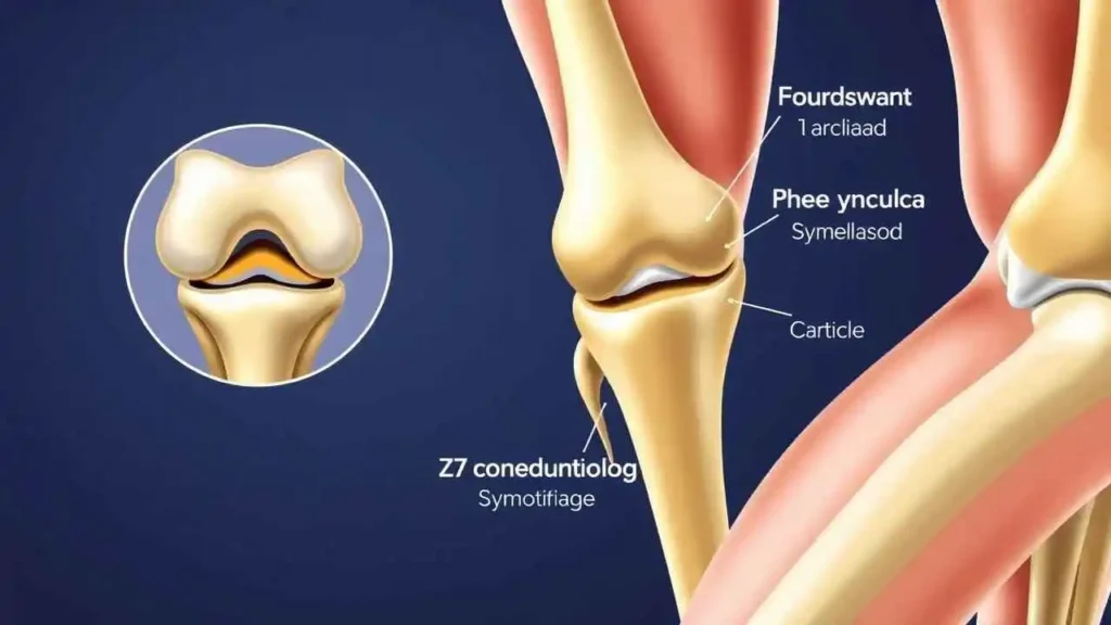
- Articular Cartilage (Hyaline Cartilage):
- Location: A smooth, pearly-white, rubbery tissue that covers the ends of the femur, the top of the tibia, and the back of the patella—essentially all areas where bones make contact within the joint.
- Composition: It is a specialized form of hyaline cartilage consisting of chondrocytes embedded in a matrix rich in collagen fibers and proteoglycans, which attract water. This high water content provides its elasticity and shock-absorbing qualities.
- Function:
- Smooth Gliding: It significantly reduces friction between bones, enabling effortless joint movement, which is one of its most impressive abilities.
- Shock Absorption: It helps distribute forces encountered during activities like walking, running, and jumping, protecting the underlying bone.
- Limitations: Articular cartilage lacks blood vessels and nerves, along with a limited capacity for cell regeneration, meaning damage to this tissue tends not to heal naturally and may progressively worsen, causing pain and functional decline.
- Menisci (Fibrocartilage):
- Location: Two crescent-shaped fibrocartilage pads situated between the femoral condyles and the tibial plateau—the medial meniscus on the inner side and the lateral meniscus on the outer side.
- Composition: Composed of tough, fibrous fibrocartilage, the menisci are more rigid and less compressible than hyaline cartilage.
- Function:
- Shock Absorption: Act as secondary shock absorbers, especially during high-impact activities.
- Load Distribution: Spread the weight transmitted from the femur over the tibial plateau, reducing concentrated stresses on the articular cartilage, much like washers distribute pressure evenly.
- Improved Fit: Their wedge-shaped structure enhances the congruency between the rounded femoral condyles and the flatter tibial plateau, increasing joint stability.
- Lubrication Assistance: Aid in circulating synovial fluid throughout the joint.
- Vulnerability: Due to their location and load-bearing role, menisci are prone to tears, particularly during twisting or rotating motions.
- Synovial Fluid:
- Location: This viscous fluid fills the cavity inside the joint capsule.
- Composition: Produced by the synovial membrane lining the capsule, it contains lubricating molecules like hyaluronic acid and essential nutrients.
- Function:
- Lubrication: Acts as a lubricant working alongside articular cartilage to minimize friction during movement, often described as the joint’s oil.
- Nourishment: Delivers nutrients and oxygen to the articular cartilage, which lacks its own blood supply, and removes metabolic waste.
- Shock Absorption: Provides a modest amount of hydrostatic cushioning within the joint.
Together, articular cartilage, menisci, and synovial fluid constitute the fundamental cushioning and lubrication system that allows the knee to operate effectively under substantial load.
Summary Table of Key Components and Their Functions
| Part | Description | Primary Function(s) |
| Bones | ||
| Femoral Condyles | Rounded ends of the thigh bone | Articulate with tibia and patella; bear body weight |
| Tibial Plateau | Flat upper surface of the shin bone | Supports femoral condyles; bears weight |
| Patella | Kneecap; embedded in quadriceps tendon | Protects the joint; enhances quadriceps leverage |
| Connective Tissue | ||
| Ligaments | Strong fibrous bands (ACL, PCL, MCL, LCL) | Provide passive joint stability; restrict excessive motion |
| Tendons | Fibrous cords connecting muscles to bones | Transmit muscular forces to enable movement |
| Cushioning/Support | ||
| Articular Cartilage | Smooth white tissue on bone ends | Reduces friction; absorbs shocks; distributes load |
| Menisci (Medial/Lateral) | C-shaped fibrocartilage pads between femur & tibia | Absorb shock; distribute load; improve joint fit and stability |
| Synovial Fluid | Viscous fluid filling joint cavity | Lubricates joint; nourishes cartilage; provides some shock absorption |
| Joint Capsule | Fibrous sac enclosing the joint | Contains synovial fluid; contributes to joint stability |
| Bursae | Small fluid-filled sacs | Reduce friction between tendons, ligaments, bones, and skin |
This comprehensive overview highlights the essential parts that work together to provide the knee with its remarkable ability to move smoothly, withstand impact, and maintain stability over a lifetime.
The Consequences of Cushion Loss
The intricate cushioning system within the knee plays a fundamental role in maintaining overall joint health. When the articular cartilage deteriorates or the menisci sustain damage, the knee’s protective barriers weaken significantly. This degradation increases friction between the bones, which often results in symptoms such as pain, inflammation, stiffness, and a grinding feel during movement. These manifestations are typical indicators of osteoarthritis, a prevalent degenerative joint disorder where the reduction of this essential cushioning layer is a major contributing factor.
The insightful quote,
“The human body is a machine that works perfectly, provided we take care of it.” — Gautama Buddha,
serves as a powerful reminder that although our knees are extraordinary feats of natural design, they are still vulnerable to the effects of aging, repetitive use, and inadequate care.
Maintaining Our Knee’s Cushioning
Because these cushioning tissues have limited capacity for repair, preserving their condition is critically important. Several strategies can help support knee health:
- Maintain a Healthy Weight: Carrying excess body weight places substantial pressure on the knee joints, hastening the degeneration of cartilage and menisci.
- Engage in Low-Impact Exercise: Activities such as swimming, cycling, and walking help to strengthen key muscles around the knee—like the quadriceps, hamstrings, and calves—without imposing undue strain on the cartilage. Robust muscles can better absorb shocks and contribute to joint stability.
- Practice Proper Technique During Exercise: Correct form when performing moves such as squats, lunges, or weightlifting is essential to minimize stress on the knees.
- Avoid Sudden, Twisting Movements: These motions commonly cause meniscal tears and ligament injuries, so steering clear reduces risk.
- Heed Your Body’s Signals: Experiencing pain is an important warning sign—never push through severe knee discomfort without seeking professional evaluation.
- Stay Hydrated and Nourished: While hydration and diet do not directly replenish cushioning, they are vital for overall tissue repair and joint function.
Understanding the anatomy and function of articular cartilage, menisci, and synovial fluid—as the knee’s natural shock absorbers—illuminates the complex and remarkable engineering of the joint alongside its susceptibility to damage. These components facilitate smooth, pain-free motion and cushion the impacts incurred daily. By recognizing the delicate balance between bones, ligaments, and protective tissues, we can be proactive in safeguarding our knees to preserve mobility for years to come. Our knees support us in countless ways throughout life; caring for their intricate cushioning system is a valuable investment in enduring health and freedom of movement.

