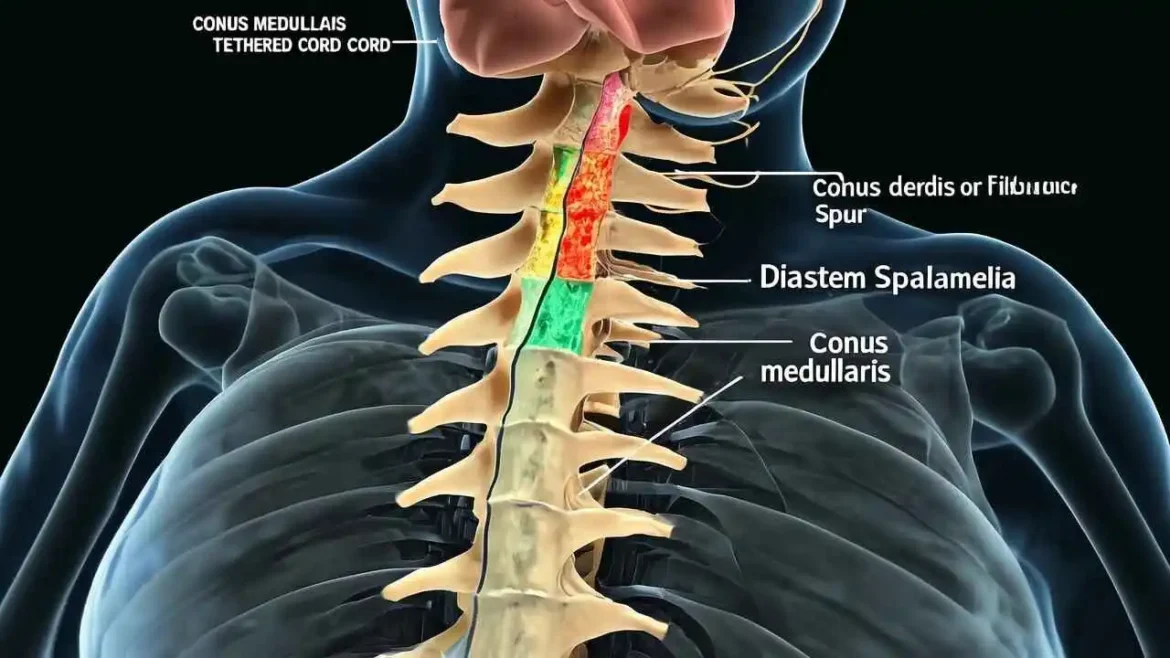
In certain situations, supplementary tests may be ordered to gather further specific information or to assess functional impact:
- Computed Tomography (CT) Scan: While MRI excels at soft tissue visualization, a CT scan may be utilized to obtain an even better, high-resolution view of the bony anatomy, particularly if there is a suspected bony septum. CT scans use X-rays to create detailed cross-sectional images of bone and calcifications. It can precisely delineate the structure of the bony spur, its dimensions, and its relationship to the surrounding vertebrae, which can be invaluable for surgical navigation when a bony resection is anticipated.
- Urodynamic Studies: If the patient is experiencing urinary symptoms, such as frequent urination, urgency, incontinence, or difficulty emptying the bladder, urodynamic studies may be ordered. The bladder and bowel are innervated by nerves originating from the lower spinal cord, and a tethered or compromised spinal cord can disrupt these nerve pathways. Urodynamic studies allow clinicians to assess bladder capacity, pressure, urine flow rates, and sphincter function. This helps to quantify the degree of bladder dysfunction, guide therapeutic interventions, and monitor the impact of the spinal cord condition on bladder health.
- Electromyography (EMG) and Nerve Conduction Studies (NCS): Although less commonly used for the initial diagnosis of Diastematomyelia itself, these neurophysiological tests may be employed to assess the function of specific nerves and muscles in the lower extremities. They can help identify whether weakness or sensory changes are due to nerve root compression or more generalized spinal cord dysfunction, and can be useful in monitoring neurological progression or recovery post-surgery.
This comprehensive diagnostic journey ensures that individuals with Diastematomyelia receive an accurate and timely diagnosis, paving the way for appropriate medical and surgical management.
Here’s an expanded version of the text, providing more detail and context for each point:
Treatment Approaches and Management of Diastematomyelia
The comprehensive management of Diastematomyelia, a rare congenital spinal malformation characterized by a longitudinal division of the spinal cord, fundamentally hinges on a crucial clinical assessment: is the patient currently symptomatic? This distinction guides the entire therapeutic strategy, balancing the risks of intervention against the potential for neurological deterioration.
Management for Asymptomatic Patients
For individuals with Diastematomyelia who are discovered incidentally – perhaps during imaging for an unrelated condition, or through routine screening for other congenital anomalies – and present with no neurological symptoms, a conservative approach of “watchful waiting” may be recommended. This strategy is predicated on avoiding unnecessary surgical risks while closely monitoring the patient’s condition.
This involves:
- Regular Neurological Check-ups: These are crucial for detecting subtle or nascent changes. Examinations typically include assessment of motor strength, sensory perception, deep tendon reflexes, gait, bladder/bowel function, and the presence of any orthopedic deformities (e.g., foot deformities, scoliosis) that might indicate spinal cord tethering.
- Periodic Imaging: Typically, repeat Magnetic Resonance Imaging (MRI) scans of the spine are performed at scheduled intervals. The primary goal is to monitor for any progression of spinal cord tethering, the development of a syrinx (a fluid-filled cyst within the cord), or changes in the bony, cartilaginous, or fibrous septum.
- Special Emphasis During Growth Spurts: Periods of rapid skeletal growth, particularly in childhood and adolescence, are critical. During these times, the spinal column lengthens significantly, potentially stretching a tethered spinal cord and exacerbating any existing tension. Monitoring during these phases is intensified due to the increased risk of symptom onset or progression.
While watchful waiting aims to prevent unwarranted surgery, it’s paramount to understand that even asymptomatic patients with clear evidence of tethering on MRI may eventually require prophylactic surgical intervention, as the risk of future neurological deficits remains high.
Management for Symptomatic Patients or Those with Radiographic Tethering
For patients who present with overt neurological symptoms, or those who, despite being asymptomatic, show clear and progressive evidence of cord tethering on MRI, surgical intervention is considered the standard of care. The rationale behind this aggressive approach is to prevent further neurological deterioration, which, once established, can be irreversible. Symptoms commonly include pain (back or leg), progressive motor weakness, sensory deficits, bladder and/or bowel dysfunction, and orthopedic deformities like scoliosis or foot deformities.
The primary goal of surgery is to prevent further neurological damage and, ideally, to induce some degree of symptom improvement.
The key objectives of the surgical procedure for Diastematomyelia are meticulously planned and executed, often employing microsurgical techniques:
- Decompression of the Neural Elements: This involves relieving any pressure exerted on the spinal cord and nerve roots. This is typically achieved through a laminectomy or laminoplasty, where part of the vertebral bone (lamina) is removed or reshaped to create more space within the spinal canal.
- Resection of the Septum: This is a critical step, involving the careful and complete removal of the abnormal midline spur. This spur can be bony (osteological), cartilaginous, or fibrous, and it directly divides the spinal cord. Its removal requires precision to avoid damaging the delicate neural tissue that often adheres to it. The type of septum (bony being the most challenging) can influence the surgical approach and complexity.
- Untethering the Spinal Cord: The Diastematomyelia septum often acts as a tether, preventing the normal caudal migration of the spinal cord as the child grows. The surgical goal is to release any adhesions, fibrous bands, or abnormal filum terminale that are pulling on the cord, allowing it to move freely within the spinal canal. This restores the cord’s physiological mobility and alleviates abnormal tension, which is the root cause of many symptoms.
- Dural Repair/Reconstruction: Diastematomyelia is classified into Type I (two separate dural sacs, each containing a hemicord) and Type II (a single dural sac containing both hemicords and the septum). If two dural sacs are present (Type I), they are often united into a single, larger sac (a procedure known as duraplasty). This reconstruction prevents the re-tethering of the cord by providing a unified, expansive space for the spinal cord to reside in and move within, and also minimizes the risk of cerebrospinal fluid (CSF) leakage. A watertight closure is essential.
Prognosis Following Surgery
The prognosis following surgical intervention for Diastematomyelia is generally good, particularly for halting the progression of symptoms and preventing further neurological decline. Many patients experience significant improvement in pain and some improvement in motor deficits, especially if the deficits were recent or mild.
However, it is crucial to manage expectations, especially for long-standing symptoms:
- Irreversible Damage: Established bladder and bowel dysfunction or significant muscle atrophy due to chronic nerve damage are less likely to be completely reversed. While some improvement may occur, full restoration of function is often challenging or impossible. This underscores the critical importance of early diagnosis and intervention before permanent and irreversible damage to the spinal cord and nerve roots occurs.
- Long-Term Follow-up: Despite successful initial surgery, there remains a lifelong risk of re-tethering, especially in growing children. Therefore, long-term neurological follow-up and periodic imaging are essential to monitor for any recurrence of symptoms or re-tethering.
- Multidisciplinary Care: Post-operative management often involves a multidisciplinary team, including physical therapists, occupational therapists, and sometimes urologists or orthopedists, to address residual deficits and optimize functional outcomes.
In summary, the management of Diastematomyelia demands a nuanced approach, prioritizing surveillance for asymptomatic cases while advocating for timely and precise surgical intervention to preserve neurological function in symptomatic individuals.
Conclusion: A Condition of Hope
Diastematomyelia represents a fascinating but serious developmental anomaly. While its origins lie in the earliest moments of life, its impact can be felt at any age. As we have seen, the journey from recognizing subtle skin markers on a baby’s back to performing complex neurosurgery is one that relies on sharp clinical suspicion and advanced technology. By understanding the causes, recognizing the diverse symptoms, and employing precise diagnostic tools, we can pave the way for effective treatment. For individuals and families facing this diagnosis, the path forward is one of proactive management and hope, aimed at protecting neurological function and ensuring the best possible quality of life.
FAQs on Preventing Diastematomyelia by Natural Remedies and Lifestyle Changes
Q1: What is diastematomyelia?
A1: Diastematomyelia is a rare congenital spinal anomaly where the spinal cord is divided into two hemicords by a median septum.
Q2: Can diastematomyelia be prevented?
A2: While there is no surefire way to entirely prevent diastematomyelia, certain natural remedies and lifestyle changes may reduce the risk.
Q3: What lifestyle changes can help prevent diastematomyelia?
A3: Maintaining a healthy diet rich in fruits, vegetables, whole grains, and omega-3 fatty acids, staying hydrated, and avoiding toxins like heavy metals and pesticides may help minimize the risk.
Q4: Are any specific nutrients crucial for preventing diastematomyelia?
A4: Folic acid, vitamin D, and certain B vitamins play key roles in spinal cord development. Ensuring adequate dietary intake or supplementing with these nutrients may be beneficial.
Q5: Can stress contribute to the development of diastematomyelia?
A5: High stress levels during pregnancy may potentially increase the risk of certain spinal cord defects, including diastematomyelia.
Q6: Are there any natural stress-relievers that could help?
A6: Techniques like meditation, deep breathing, and yoga have stress-reducing effects and may be beneficial for expectant mothers.
Q7: How does physical exercise impact the risk of diastematomyelia?
A7: Regular exercise, especially during pregnancy, may promote healthy spinal development and potentially lower the risk of certain spinal anomalies.
Q8: Are there any specific exercises recommended for expectant mothers?
A8: Gentle exercises like walking, swimming, and prenatal yoga can be safe and beneficial for overall health during pregnancy.
Q9: Can poor maternal nutrition increase the risk of diastematomyelia?
A9: Yes, inadequate maternal nutrition, particularly a deficiency in essential vitamins and minerals, may contribute to an elevated risk of congenital spinal anomalies.
Q10: What vitamins and minerals are crucial for fetal spinal development?
A10: Folic acid, vitamin B12, vitamin D, and calcium are essential for normal fetal growth and spinal cord development.
Q11: Are there any natural remedies that could enhance spinal health during pregnancy?
A11: Adding natural sources of omega-3 fatty acids (like fish oil), glucosamine, and chondroitin to one’s diet may support spinal health.
Q12: Can a prenatal vitamin with adequate nutrients help protect against diastematomyelia?
A12: A well-formulated prenatal vitamin that includes essential vitamins and minerals may help support fetal spinal development and potentially lower the risk.
Q13: How does a mother’s weight during pregnancy influence the risk of diastematomyelia?
A13: Excessive maternal weight gain during pregnancy may increase the risk of certain spinal anomalies, including diastematomyelia.
Q14: Are there any specific dietary recommendations for expectant mothers to minimize the risk of diastematomyelia?
A14: A balanced diet that includes lean proteins, whole grains, fruits, and vegetables, with moderate amounts of healthy fats, can support overall health during pregnancy.
Q15: Can environmental toxins like pesticides increase the risk of diastematomyelia?
A15: Exposure to certain environmental toxins, such as pesticides, may increase the risk of congenital spinal anomalies, though the exact relationship is not fully understood.
Q16: Are there any natural ways to reduce exposure to environmental toxins?
A16: Maintaining a clean living environment, avoiding certain chemicals, and using natural personal care products can help minimize exposure to potential toxins.
Q17: How does folic acid supplementation during pregnancy impact diastematomyelia risk?
A17: Adequate folic acid intake before and during early pregnancy has been shown to significantly reduce the risk of certain spinal cord defects, including diastematomyelia.
Q18: Can other B vitamins also play a role in preventing diastematomyelia?
A18: While folic acid is most closely associated with preventing neural tube defects, other B vitamins, like B12 and B9, also contribute to fetal growth and spinal cord development.
Q19: Are there any risks associated with high doses of folate or other B vitamins during pregnancy?
A19: While B vitamins are essential, excessive intake can lead to adverse effects, so it is crucial to follow recommended prenatal supplement guidelines.
Q20: Can certain food additives increase the risk of diastematomyelia?
A20: While the link between food additives and diastematomyelia is not well established, a diet rich in whole foods and avoiding processed items may be a sensible approach to minimizing overall health risks.
Q21: Are there any natural remedies that could alleviate symptoms if a child is born with diastematomyelia?
A21: While there is no cure for diastematomyelia, therapies like physical therapy, bracing, and possibly surgery can help manage symptoms and improve quality of life.
Q22: Can lifestyle changes and natural remedies alone effectively manage diastematomyelia?
A22: While lifestyle modifications and natural remedies may be beneficial for overall health, they are unlikely to fully manage the symptoms of diastematomyelia, which often requires a combination of medical interventions and therapies.
Q23: When should a pregnant woman consult a healthcare provider about diastematomyelia risk?
A23: Anytime concerns arise about congenital anomalies, a healthcare provider should be consulted. This includes before becoming pregnant, during early pregnancy, and if there is a family history of spinal cord defects.
Q24: What prenatal tests can detect diastematomyelia?
A24: While diastematomyelia is often diagnosed postnatally through imaging tests like MRI, certain prenatal screenings like ultrasound may identify some cases, although accuracy can vary.
Q25: Can a healthy diet and lifestyle reduce the risk of other congenital anomalies as well?
A25: Yes, maintaining a healthy diet and lifestyle during pregnancy can help minimize the risk of various congenital anomalies, not just diastematomyelia.
Q26: How does maternal age impact the risk of diastematomyelia?
A26: The risk of certain congenital anomalies, including diastematomyelia, increases with advancing maternal age, especially after 35 years old.
Q27: Are there any natural ways to support healthy fetal development in older mothers?
A27: A well-balanced diet, staying physically active, and managing stress can all support healthy fetal development, even for older mothers.
Q28: Can certain medications increase the risk of diastematomyelia?
A28: While most medications are safe during pregnancy, certain drugs like isotretinoin (Accutane) are teratogenic and should be avoided due to their potential to cause severe birth defects.
Q29: What prenatal supplements are recommended to minimize the risk of diastematomyelia?
A29: Taking a well-formulated prenatal vitamin with folic acid, vitamin D, and other essential nutrients is recommended to support fetal development and potentially lower the risk of congenital anomalies.
Q30: Are there any ongoing research studies exploring natural prevention strategies for diastematomyelia?
A30: Researchers are continually investigating the relationships between various risk factors and congenital anomalies, including diastematomyelia. While no conclusive evidence supports natural prevention strategies at this time, ongoing studies may uncover new insights in the future.
Medical Disclaimer:
The information provided on this website is for general educational and informational purposes only and is not intended as a substitute for professional medical advice, diagnosis, or treatment. Always seek the advice of your physician or other qualified health provider with any questions you may have regarding a medical condition. Never disregard professional medical advice or delay in seeking it because of something you have read on this website.


