Hematemesis: Why You Should Never Ignore Vomiting Blood
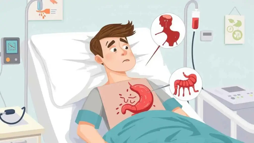
Experiencing or witnessing someone vomit blood is profoundly alarming. It’s a symptom that immediately signals something is significantly wrong within the body. Medically known as hematemesis, this isn’t just general sickness; it’s the forceful expulsion of blood from the stomach, typically originating from the upper gastrointestinal (GI) tract. As we delve into this serious topic, our goal is to provide a clear, informative overview of hematemesis, exploring its definition, common causes, recognizable symptoms, how we approach diagnosis, and the crucial steps involved in treatment. We understand the fear and confusion this symptom can bring, and our aim is to shed light on its various facets.
What Exactly is Hematemesis?
At its core, hematemesis is the act of vomiting blood. It’s distinct from hemoptysis, which is coughing up blood from the respiratory tract (lungs and airways). With hematemesis, the blood originates from structures higher up in our digestive system – primarily the esophagus (the tube connecting the mouth to the stomach), the stomach itself, or the duodenum (the first part of the small intestine).
The appearance of the vomited blood can vary widely, and this variation often provides us with critical clues about the source and duration of the bleeding. Bright red, fresh blood suggests rapid, active bleeding, possibly from an artery or a significant vein, often higher up in the esophagus or stomach. Darker blood or material resembling coffee grounds indicates that the blood has been sitting in the stomach for some time, where it has been exposed to acidic gastric juices. This exposure causes the hemoglobin in the blood to oxidize, changing its color and texture. Sometimes, the vomit might contain both fresh blood and coffee-ground material.
For us, recognizing hematemesis is the first vital step, understanding that it’s a medical emergency requiring immediate attention.
Recognizing Hematemesis: The Symptoms We Look For
The most obvious symptom of hematemesis is, of course, the vomiting of blood. However, we often see this accompanied by other signs and symptoms, which together can help us understand the severity and potential cause of the bleeding.
Common symptoms we might observe alongside hematemesis include:
- Nausea: A feeling of sickness or urge to vomit often precedes or accompanies the event.
- Weakness or Fatigue: Significant blood loss can lead to anemia, causing generalized weakness.
- Dizziness or Lightheadedness: Reduced blood volume and pressure can cause these symptoms, especially when standing up quickly.
- Pale Skin (Pallor): Another sign of potential blood loss and anemia.
- Shortness of breath (Dyspnea): Especially if blood loss is substantial.
- Abdominal Pain or Discomfort: Depending on the underlying cause, such as ulcers or inflammation.
- Tachycardia (Rapid Heart Rate): The heart pumps faster to compensate for reduced blood volume.
- Hypotension (Low Blood Pressure): In cases of significant or rapid blood loss.
- Melena: Black, tarry stools. This indicates that blood has passed through the digestive tract and been altered by bacteria in the intestines. It’s a sign of bleeding further up the GI tract.
When we see hematemesis combined with signs of shock (very low blood pressure, rapid heart rate, cold clammy skin), we know the situation is critical and immediate medical intervention is paramount.
Why Does Hematemesis Occur? Exploring the Causes
Hematemesis isn’t a condition itself, but rather a symptom stemming from underlying issues causing bleeding in the upper GI tract. The list of potential causes is extensive, ranging from common and relatively benign issues to severe, life-threatening conditions. Understanding these causes is crucial for us to make an accurate diagnosis and plan effective treatment.
Here are some of the most frequent causes we encounter:
- Peptic Ulcers: These are open sores that develop on the lining of the stomach or the first part of the small intestine (duodenum). They are often caused by Helicobacter pylori infection or the use of nonsteroidal anti-inflammatory drugs (NSAIDs) like ibuprofen or aspirin. Bleeding occurs when the ulcer erodes into a blood vessel.
- Esophageal Varices: These are enlarged, swollen veins in the lining of the lower esophagus, often a complication of severe liver disease (like cirrhosis). Increased pressure in the portal vein system (portal hypertension) causes these veins to bulge. They are fragile and prone to rupture, leading to massive, often sudden and severe, bleeding.
- Mallory-Weiss Tear: This is a tear in the mucous membrane, or lining, of the esophagus, where it joins the stomach. It’s typically caused by forceful vomiting or retching, often associated with binge drinking. While usually not as severe as variceal bleeding, it can still cause significant blood loss.
- Esophagitis or Gastritis: Inflammation of the esophagus (esophagitis) or stomach (gastritis) can erode the lining, leading to superficial bleeding. Causes include excessive alcohol consumption, chronic vomiting, certain medications, or acid reflux (GERD).
- Cancers: Cancers of the esophagus or stomach can cause bleeding as the tumor grows and erodes into blood vessels.
- Vascular Malformations: Abnormal or weakened blood vessels (like angiodysplasia or Dieulafoy’s lesion) can be present in the upper GI tract lining and are prone to bleeding.
- Ingestion of Caustic Substances: Swallowing corrosive chemicals can severely damage the lining of the esophagus and stomach, leading to bleeding.
To help organize some of the key causes, we can look at this table:
| Common Cause | Brief Description | Severity |
| Peptic Ulcers | Open sores in stomach or duodenum lining, often due to H. pylori or NSAIDs. | Can range from mild ooze to severe arterial bleed |
| Esophageal Varices | Enlarged veins in lower esophagus, due to liver disease (portal hypertension). | High risk of massive, life-threatening bleed |
| Mallory-Weiss Tear | Tear in esophageal lining at stomach junction, usually from forceful vomiting. | Often self-limiting, but can bleed significantly |
| Esophagitis/Gastritis | Inflammation of esophagus/stomach lining, due to acid, alcohol, meds, etc. | Usually mild bleeding, but can be persistent |
| Cancers | Malignant tumors in esophagus or stomach. | Variable, can cause significant erosion/bleed |
| Vascular Malformations | Abnormal blood vessels in lining (e.g., angiodysplasia). | Can cause intermittent or chronic bleeding |
We approach identifying the cause by considering the patient’s medical history, lifestyle, medication use, and examining the characteristics of the vomit.
Diagnosing the Source: How We Investigate
When a patient presents with hematemesis, our first priority is to stabilize them. This involves assessing their vital signs (blood pressure, heart rate, breathing) and ensuring they are not in shock. We initiate intravenous fluids and, if necessary, blood transfusions to replace lost volume.
Once the patient is stable, our focus shifts to pinpointing the exact location and cause of the bleeding. This involves:
- Taking a Detailed History: We ask about the appearance and amount of blood, how long it’s been happening, any prior GI issues (ulcers, liver disease), medication use (especially NSAIDs, blood thinners), alcohol consumption, and recent episodes of vomiting.
- Physical Examination: We check for signs of anemia, liver disease (jaundice, swollen abdomen), abdominal tenderness, and signs of shock.
- Laboratory Tests:
- Complete Blood Count (CBC): To assess the level of anemia and platelet count.
- Coagulation Studies: To check if the blood is clotting properly, which can be affected by liver disease or medications.
- Liver Function Tests: To assess for underlying liver disease.
- Blood Type and Crossmatch: If a blood transfusion is needed.
- Endoscopy (Esophagogastroduodenoscopy – EGD): This is the cornerstone of diagnosing upper GI bleeding. We insert a thin, flexible tube with a camera down the esophagus, into the stomach, and the first part of the duodenum. This allows us to directly visualize the lining, identify the bleeding source (ulcer, varix, tear, etc.), and often, treat it simultaneously. We typically perform an EGD as soon as the patient is stable.
- Other Imaging Studies: Less commonly used for active bleeding compared to endoscopy, but imaging like CT angiography might be used if endoscopy is unsuccessful or the bleeding source is suspected to be unusual or deeper.
Based on these diagnostic steps, we can usually identify the cause and severity of the bleeding, guiding our treatment strategy.
Treating Hematemesis: Stopping the Bleed and Addressing the Cause
Treating hematemesis involves two main objectives: first, stopping the active bleeding and stabilizing the patient, and second, treating the underlying cause to prevent future episodes.
Our treatment approach is often multi-faceted:
- Resuscitation and Stabilization: This is always the immediate priority. We administer IV fluids to maintain blood pressure and adequate circulation. Blood transfusions are given if the patient has significant blood loss or anemia.
- Endoscopic Hemostasis: Once the bleeding source is identified during EGD, we use various techniques delivered through the endoscope to stop it. These include:
- Injection: Injecting a substance (like epinephrine or saline) directly into or around the bleeding site to constrict blood vessels.
- Thermal Therapy: Using heat (cautery) or cold (cryotherapy) probes to seal the bleeding vessel.
- Mechanical Methods: Applying clips or bands directly onto the bleeding vessel or tissue to stop the flow. This is particularly common for esophageal varices (banding).
- Medications:
- Proton Pump Inhibitors (PPIs): These powerful acid-reducing drugs (like omeprazole, pantoprazole) are essential for bleeding caused by ulcers or erosions. They help stop bleeding and promote healing.
- Vasoactive Drugs: Medications like octreotide or vasopressin are used specifically for bleeding esophageal varices. They help reduce pressure in the portal vein system, thereby reducing blood flow to the varices.
- Antibiotics: If H. pylori infection is the cause of a peptic ulcer, we prescribe antibiotics.
- Interventional Radiology: In some cases, particularly for severe bleeding that can’t be controlled endoscopically, interventional radiologists can perform procedures like angiography with embolization, where they thread a catheter through blood vessels and inject material to block the bleeding artery.
- Surgery: Surgery is usually a last resort, typically needed for severe, life-threatening bleeding that doesn’t respond to endoscopic or radiological interventions, or for complex cases like perforated ulcers or certain tumors.
- Treating the Underlying Cause: Once the acute bleeding is controlled, we focus on managing the condition that caused it. This might involve long-term medication for ulcers, managing liver disease, addressing alcohol use, or treating cancer.
As the great physician Sir William Osler wisely stated:
“The good physician treats the disease; the great physician treats the patient who has the disease.”
This quote reminds us that while we focus on the urgent task of stopping the bleeding, we also treat the whole person, considering their underlying health, lifestyle, and the impact of this event on their life.
Why Hematemesis Can Be Crucial and What Precautions Can Be Taken
Hematemesis, the vomiting of blood originating from the upper gastrointestinal (GI) tract—typically the esophagus, stomach, or duodenum—is a serious medical symptom that requires immediate attention. The presence of blood in vomit indicates internal bleeding, which can quickly escalate into a life-threatening condition if not addressed promptly.
Why Hematemesis Is Crucial
The severity of hematemesis depends on the volume and rate of bleeding. Even small amounts of blood over time can lead to anemia, while massive bleeding can cause hypovolemic shock—a dangerous drop in blood volume that impairs oxygen delivery to tissues. This can result in dizziness, rapid heartbeat, low blood pressure, confusion, and even death if not treated urgently.
Precautions to Take
When dealing with hematemesis, certain precautions can help manage the situation effectively and prevent further deterioration:
Seek Immediate Medical Help : Hematemesis should always be considered a medical emergency. Call for professional assistance or go to the nearest emergency room without delay.
Do Not Eat or Drink : Until evaluated by a doctor, avoid consuming food or beverages, especially alcohol or caffeine, which can worsen GI irritation.
Avoid Medications That Irritate the Stomach : Refrain from taking nonsteroidal anti-inflammatory drugs (NSAIDs) like ibuprofen or aspirin unless prescribed, as they can increase gastric bleeding.
Stay Calm and Rest : Stress and physical exertion can elevate heart rate and blood pressure, potentially worsening bleeding. Lie down and stay calm until help arrives.
Monitor Symptoms : Keep track of how much blood was vomited, its color (bright red vs. coffee-ground), and any associated symptoms like dizziness, chest pain, or fainting. This information is valuable for healthcare providers.
Avoid Alcohol and Tobacco : Both substances can exacerbate conditions like gastritis, ulcers, and liver disease, increasing the risk of recurrent bleeding.
Follow Medical Advice Post-Treatment : After diagnosis and treatment, strictly follow your doctor’s recommendations regarding medications, diet, and lifestyle changes to prevent recurrence.
Manage Underlying Conditions : If hematemesis is linked to chronic issues like GERD, peptic ulcer disease, or liver cirrhosis, regular medical follow-ups and proper management are essential.
Maintain a Healthy Diet : Avoid spicy, acidic, or overly processed foods that can irritate the stomach lining. Opt for bland, easily digestible meals if recovering from GI bleeding.
Control Liver Disease Risk Factors : For individuals with cirrhosis, managing alcohol intake, maintaining a healthy weight, and treating hepatitis infections are critical steps in preventing esophageal varices and related complications.
Understanding its significance and taking appropriate precautions can make a significant difference in outcomes. Prompt intervention, accurate diagnosis, and consistent follow-up care are key to managing this condition and preventing future episodes.
How to Differentiate Between Hemoptysis and Hematemesis